Autism marker spied in brain folds
 Researchers say they have found a brain marker for autism that can be detected by MRI and is present as from the age of two.
The abnormality is a less deep fold in Broca's area - a region of the brain linked to language and communication; functions that are impaired in autistic patients.
The autistic spectrum covers a range of neuro-developmental disorders that mainly affect social relationships and communication.
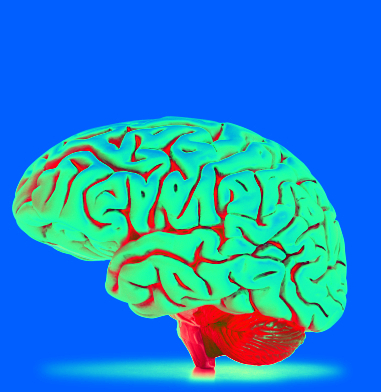
Recent neuroimaging research suggests abnormal cortical folding (the formation of convolutions on the surface of the brain) is involved in the development of autism, but until now markers specific to the different kinds of disorders had not been pinpointed.
Scientists at the Institut de Neurosciences de la Timone (in Marseille, France) took the search to the “sulcal pit” - the deepest point of each sulcus in the cerebral cortex, from which points all the folds on the brain's surface develop.
It is believed that this area forms at a very early developmental stage, probably under genetic influences, and so is useful for comparisons between different individuals.
The researchers observed (via MRI) the sulcal pits of 102 boys aged 2 to 10 years in three groups: those with autistic disorder, pervasive developmental disorder not otherwise specified, and typically developing children.
The comparison showed that in Broca's area (a region known to be involved in language and communication), the maximum depth of a sulcus was less among autistic children than for the other two groups.
Interestingly, the team found that the atrophy was linked with the social communication performance of children in the autistic group - the deeper the sulcal pits, the more impaired their skills in language production were.
The team said the abnormality specific to autistic children can be used as a biomarker that could enable their earlier diagnosis and management, as early as from the age of two years.


 Print
Print