Nervous COVID effect shown
 A new study reveals SARS-CoV-2 rapidly infects the nervous system prior to bloodstream entry.
Researchers have uncovered critical insights into how SARS-CoV-2, the virus responsible for COVID-19, works within the body.
The virus is shown to rapidly infect the nervous system before entering the bloodstream. This infection occurs through peripheral nerves connected to sensory and autonomic systems, leading to significant damage in these neurons.
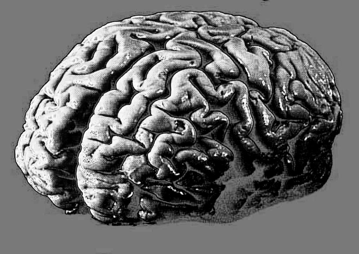
The virus was detected in various brain regions within days of infection, indicating that it can invade the central nervous system via nerve pathways rather than through the bloodstream.
The findings have significant implications for understanding the neurological impacts of COVID-19, including its association with Long COVID.
The study examined the neuronal infection of SARS-CoV-2 across various animal models and neuronal cell cultures.
Researchers aimed to determine if infection of the peripheral nervous system (PNS) preceded and contributed to central nervous system (CNS) infection.
They conducted experiments using wild-type (WT) mice and human ACE2-expressing mice (hACE2), exposing them to a 2020 variant of SARS-CoV-2 via intranasal inoculation.
Diagnostic tests revealed low levels of blood and lung infection in both groups at 3 and 6 days post-infection.
However, the virus was found in significant concentrations in the trigeminal ganglia (TG) and superior cervical ganglia (SCG), with active viral genome replication observed in both hACE2 and WT mice.
The study highlighted severe pathology in hACE2 mice, with about 97 per cent of neurons in the SCG showing substantial damage.
The viral nucleocapsid (N) protein was present in these neurons, leading to a loss of ganglionic architecture.
Notably, viral RNA was also found in various brain regions, including the olfactory bulb, hippocampus, cortex, brainstem, and cerebellum, by 3 days post-infection, increasing further at 6 days post-infection.
The researchers found that SARS-CoV-2 could infect PNS sensory neurons and functionally connected CNS tissues, suggesting that the virus can invade the CNS through axonal transport rather than through blood entry.
This was evidenced by the detection of viral RNA in PNS ganglia and connected CNS tissues within 18 hours post-infection, long before any viral RNA was found in the bloodstream.
The study also explored the role of neuropilin-1 (NRP-1) as an alternative receptor for SARS-CoV-2 entry into neurons, providing insights into how the virus can infect non-ACE2 expressing cells.
These findings show the complexity of COVID-19’s neurological impact.
Neurological symptoms are common in COVID-19 patients, with many experiencing long-term effects such as fatigue, memory issues, and ‘brain fog’.
The study concludes that neuroinvasion is not a rare event following SARS-CoV-2 infection and occurs well before the onset of symptomatic disease.
As COVID-19 transitions to an endemic disease, understanding its long-term neurological effects becomes crucial.
The study's approach ties together various lines of research, providing a clearer picture of how SARS-CoV-2 affects the nervous system.
Experts say ongoing research into the virus’s mechanisms of infection and its long-term neurological consequences is needed.


 Print
Print